Researchers from Tohoku University in Japan have Developed a Lightweight Deep Learning Model for Automatic Segmentation and Analysis of Ophthalmic Images
The human eye is said to act as a window for assessing general health. Similar to cerebral and coronary microcirculation in terms of anatomy and physiology is the retinal microvasculature. Therefore, knowledge of retinal microvascular structure and changes through time can help doctors identify and treat ocular, cardiovascular, and brain diseases.
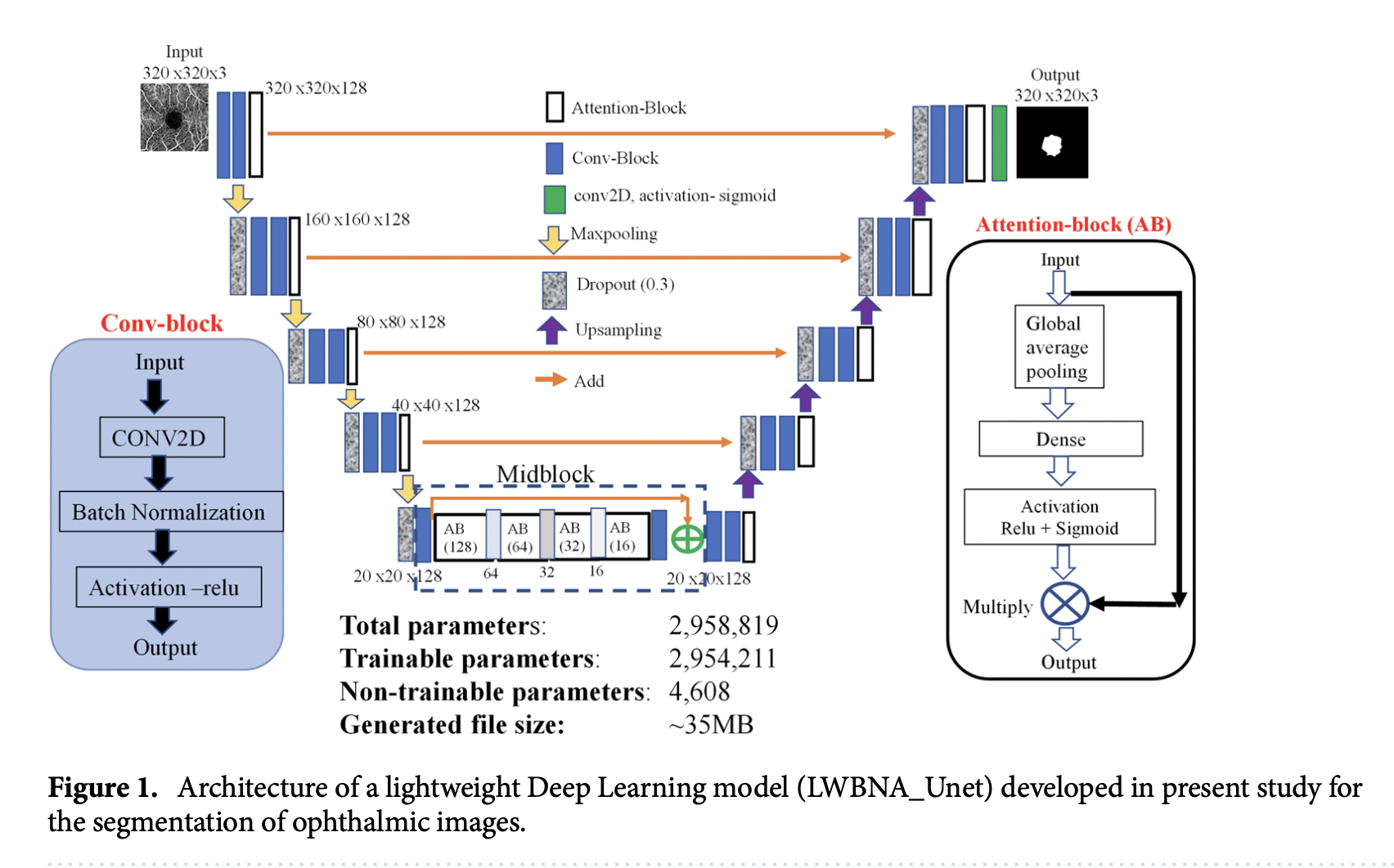
Researchers from Tohoku University in Japan have developed a lightweight deep learning model (LWBNA_Unet) for automatic segmentation and analysis of ophthalmic images. The so-called “lightweight” deep learning (DL) model is 10 times lighter than the most popular model (Unet) in biomedical image segmentation and may be applied to mobile devices and trained using just a few photos, even ones with a lot of noise.
Accurately assessing tumors, tissue volume, or other types of anomalies is necessary for diagnosing disorders. To do this, a model must examine distinct pictures and segment them by drawing lines along edges. Since accurate prediction requires more processing power, it is challenging to implement them on mobile devices.
The researchers claim that while tele-screening for illnesses and self-monitoring based on DL models are becoming more commonplace, deep learning algorithms are often task-specific, recognizing or detecting broad objects like people, animals, or traffic signs.

Regarding deep learning DL models, accuracy, speed, and processing resources are always trade-offs. The proposed model is more effective and lightweight when compared to other commercial software since it has improved segmentation accuracy and model training repeatability, even with fewer parameters. The” model is also capable of detecting/segmenting optic discs and hemorrhages in fundus pictures with great precision.” Without the requirement for a bigger training dataset, the model may be taught to accurately segment a range of ophthalmic pictures as well as to classify illnesses.
To improve glaucoma screening, scientists took measurements of the foveal avascular zone—an area containing the fovea centralis at the retina’s center—using low-resource instruments.
The team intends to use the portable model in the future to check for various illnesses and other common eye conditions.
This Article is written as a summary article by Marktechpost Staff based on the paper  'A lightweight deep learning model for automatic segmentation and analysis of ophthalmic images'. All Credit For This Research Goes To Researchers on This Project. Checkout the paper and reference article.

Please Don't Forget To Join Our ML Subreddit
Credit: Source link
Comments are closed.