Latest Artificial Intelligence Research Presents Generative Adversarial Neural Networks Capable of Generating DeepFake Knee Osteoarthritis X-rays with Varying Osteoarthritis Severity
Recent advances in deep learning have touched the field of medical science. However, recent privacy concerns and legislative frameworks have hampered the sharing and acquisition of medical data. Such legislative restrictions restrict the potential for future advancements in deep learning, which is a particularly data-intensive technique and partnership. However, producing synthetic data that is accurate for medical purposes can reduce privacy concerns and improve deep learning pipelines. This paper introduces generative adversarial neural networks that can have accurate images of X-rays of knee joints with varying degrees of osteoarthritis. Researchers provide 5,556 genuine photos along with 320,000 artificial (DeepFake) X-ray images for training.
With the help of 15 medical professionals, they evaluated our models for medical accuracy and looked at the effects of augmentation on a task that classified the severity of osteoarthritis. For medical professionals, they created a survey using 30 actual and 30 DeepFake photos. As a result, more DeepFakes than the opposite were often taken for the real thing. The outcome indicated that DeepFake realism was sufficient to fool medical professionals. Finally, using limited real data and transfer learning, our DeepFakes increased classification accuracy in a challenge to classify the severity of osteoarthritis. Additionally, they substituted all genuine training data in the same classification job with DeepFakes, and the accuracy of categorizing true osteoarthritis X-rays suffered only a 3.79% loss from baseline.
Early detection can slow the clinical course and potentially enhance the patient’s mobility and quality of life. Medical professionals, as well as artificial neural networks, have substantial difficulties in early diagnosis. With the help of two generative adversarial neural networks, they were able to create an infinite number of knee osteoarthritis X-rays at various Kellgren and Lawrence stages for this investigation. Researchers first demonstrated anonymity and augmentation effects in deep learning, and then researchers validated their system with 15 medical professionals. The generated DeepFake X-ray images can be freely shared among researchers and members of the public.
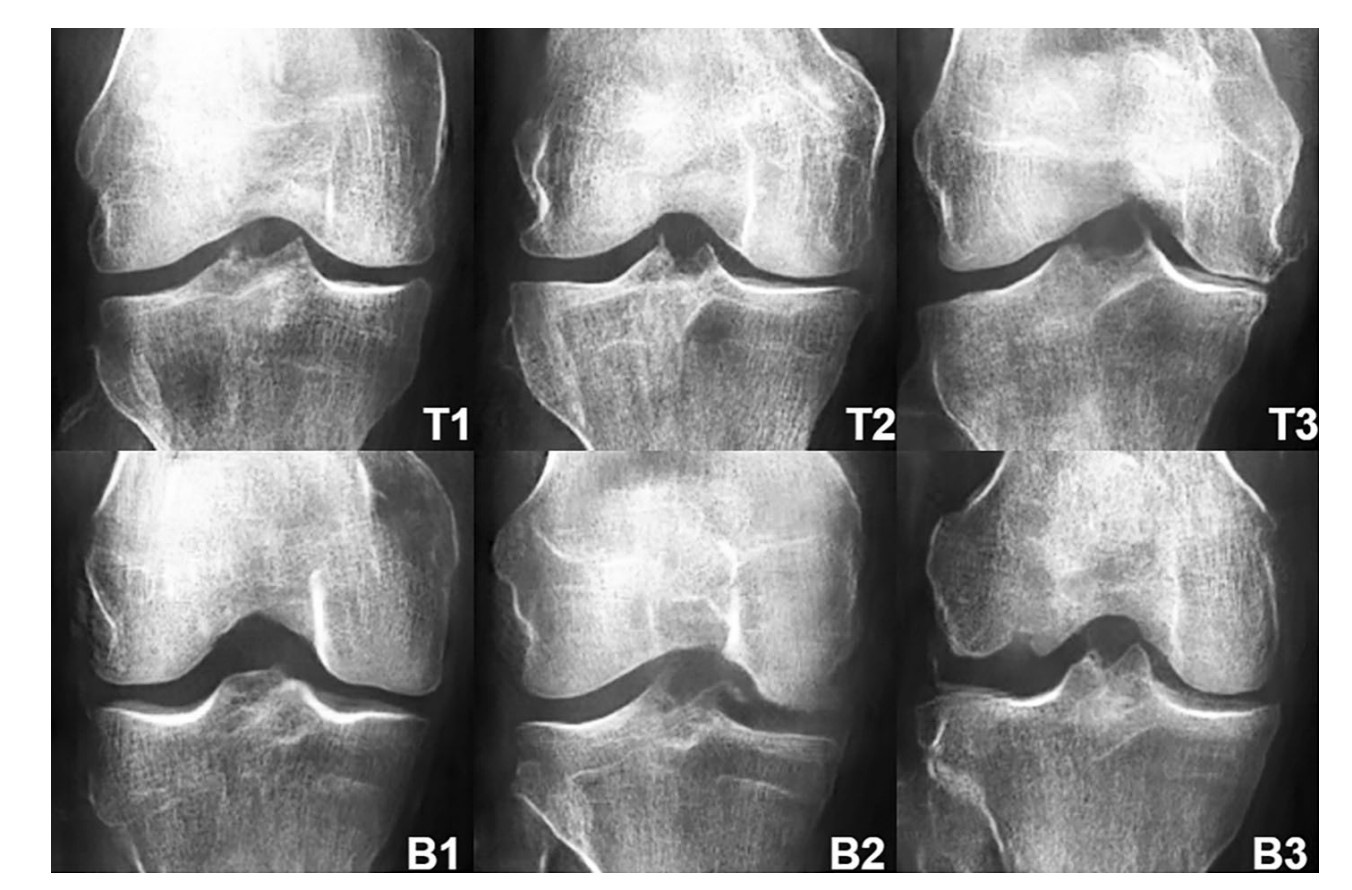
The photos for KL01 WGAN and KL234 WGAN ranged from early training up to the best-selected models.
On X-ray pictures of the human anatomy, neural networks for KL01 WGAN and KL234 WGAN were trained. As the training went on, they noticed that significant structural changes started to lessen while texture modifications improved. Upsampling and 2D convolution modules with exponential unit activations and batch normalization were the main building blocks used to construct the generator block. The dropout layers to prevent overfitting made the discriminator block a unique analysis of 30 authentic and 30 fake DeepFake photos from the KL01 and KL234 classes. The degree of OA was rated by experts for both genuine and artificial images. Results showed that more bogus photos than actual ones were mistaken for one another. Between KL01 and KL234 OA severities were predicted using the binary classification task.
For the DeepFake augmentation set, researchers saw that losses were decreased, and validation accuracy increased as a result. The augmentation effect with the highest testing score, +200% Fakes, was the most effective. Overall, both amplification and anonymization effects suggested the possibility of beneficial downstream consequences in the classification of knee osteoarthritis. Deep neural networks may be able to produce X-rays of knee osteoarthritis that are medically accurate. The linked amplification effects and anonymity by replacement were first obtained in this study.
In order to increase classification accuracy in transfer learning with limited data, DeepFake images were added to actual training data. Such transfer learning strategies are widely used in the medical field, where data are frequently in short supply and challenging to gather. To prevent GPU memory overflow, an image size of 210 x 210 was employed. To increase the number of photos available for two models of osteoarthritis severity, they combined KL classes (KL01 and KL234). Early KL grades experienced less label noise as a result of the combination of KL grades.
Focus filtering was employed to prevent focused and unfocused textures from being combined into one image since large gaps in X-ray focus and texture clarity would confuse the generator. To distinguish DeepFake images from real photos, experts needed assistance. The substantial standard deviations seen in the KL rating agreement task also reflect the presence of this effect. The assessments of medical professionals were skewed since some photos showed superior clinical attributes than others. The production and detection of landmarks may benefit from further integration of landmark labels.
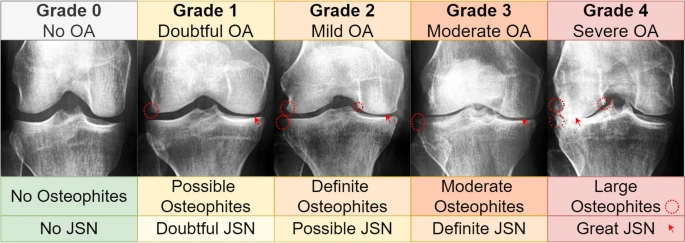
The 4130 X-rays that included both knee joints were used to create the images, which were then graded using the Kellgren and Lawrence system. There were 3253 photos for grade zero, 1495 for grade one, 2175 for grade two, 1086 for grade three, and 251 for grade four in the KL. The study’s goal was to look into how realistic DeepFake photos are. They generated 15 KL01 and 15 KL234 photos at random and then asked medical professionals to judge them based on their KL scores.
Images were resized to 315 315 pixels and included to the survey in a random sequence. They used the balanced accuracy metric79 to deal with unbalanced responses. The study team employed a straightforward variation of the ImageNet-pretrained VGG1664 architecture that was further trained for 22 epochs, with only the final three blocks of the design being trainable and the rest being frozen. To generate each dataset, they started with actual data and gradually added more DeepFake data. Using the Python language’s “random” package, real photos were chosen at random.
Check out the Paper and Dataset. All Credit For This Research Goes To Researchers on This Project. Also, don’t forget to join our Reddit page and discord channel, where we share the latest AI research news, cool AI projects, and more.
Prezja, F., Paloneva, J., Pölönen, I. et al. DeepFake knee osteoarthritis X-rays from generative adversarial neural networks deceive medical experts and offer augmentation potential to automatic classification. Sci Rep 12, 18573 (2022). https://doi.org/10.1038/s41598-022-23081-4
![]()
Ashish kumar is a consulting intern at MarktechPost. He is currently pursuing his Btech from the Indian Institute of technology(IIT),kanpur. He is passionate about exploring the new advancements in technologies and their real life application.
Credit: Source link
Comments are closed.